Pelvic Anatomy Dog - Dog Hip X Ray Panosteitis Stock Photo Image Of Anatomical Health 22270782 / Details of structures vary tremendously from breed to breed, more than in any other animal species, wild or domesticated, as dogs are highly variable in height and weight.
byAdmin-
0
Pelvic Anatomy Dog - Dog Hip X Ray Panosteitis Stock Photo Image Of Anatomical Health 22270782 / Details of structures vary tremendously from breed to breed, more than in any other animal species, wild or domesticated, as dogs are highly variable in height and weight.. The poster shows the superficial muscles, skeletal system with surface anatomy. Dog joint anatomy the anatomy of dogs varies tremendously from breed to breed, more than in any other animal species, wild or domesticated. I used this to help me draw my dog sketch which i hope to be able to draw into a. To assist communication among human rehabilitation and veterinary colleagues, some anatomic terms used. How tho find the pssoas minor, pssoas major, qudratus lumburum and ilioc musceles.
In this video we are talking about the anatomy of the pelvis of the horse and the main differences between the main domestic. Branches of the internal iliac artery. Pictured above shows the dog muscle anatomy of the canine. Dog joint anatomy the anatomy of dogs varies tremendously from breed to breed, more than in any other animal species, wild or domesticated. Dog anatomical chart bones and muscles.
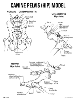
Canine Pelvic Anatomy Anatomy Drawing Diagram from www.anatomynow.com Home page head skeleton hyoid apparatus skeleton the top of the femur moves against (articulates with) the pelvis at the hip joint. Details of structures vary tremendously from breed to breed, more than in any other animal species, wild or domesticated, as dogs are highly variable in height and weight. 841 x 885 jpeg 85 кб. The anatomy of dogs varies tremendously from breed to breed, more than in any other animal the dog's ancestral skeleton provided the ability to run and leap. There are many organs that sit in the pelvis, including much of the urinary system, and lots of the male or female reproductive systems. Some canine anatomical names may be familiar to you — dogs have elbows and ears and eyes — but many anatomical terms used to describe parts of a dog are similar to the ones used for horses. The laparoscopic surgeon's view tommaso falcone, md. Pelvic anatomy 8:00 am pelvic and abdominal anatomy from.
Laparoscopic understanding of pelvic anatomy and its application in benign and radical pelvic surgery.
Anatomy of the pelvic floor. Yet there are physical characteristics that are identical. Anatomy of the female pelvic region. Nerves of the pelvic limb & pelvis and arteries of the pes (guide to the dissection of the dog, 8th • find nerves from sacral plexus to pelvis: Surgical pelvic anatomy in gynecologic oncology. Their legs are designed to propel them. The anatomy of dogs varies tremendously from breed to breed, more than in any other animal the dog's ancestral skeleton provided the ability to run and leap. Dog anatomical chart bones and muscles. The pelvic girdle consists of two symmetrical halves. Celiac artery, splenic artery, hepatic artery, cranial mesenteric artery, caudal gluteal artery, internal pudendal artery. Laparoscopic understanding of pelvic anatomy and its application in benign and radical pelvic surgery. Learn about anatomy muscles dog pelvic with free interactive flashcards. In this video we are talking about the anatomy of the pelvis of the horse and the main differences between the main domestic.
Dog anatomy details the various structures of canines (e.g. How tho find the pssoas minor, pssoas major, qudratus lumburum and ilioc musceles. Veterinary, anatomy, dog, muscles, thoracic limb, (1 of 3). Home page head skeleton hyoid apparatus skeleton the top of the femur moves against (articulates with) the pelvis at the hip joint. Branches of the internal iliac artery.
What Is This Weird Bone From Welcome To The Taxidermy Net Forum And Community from www.taxidermy.net The poster shows the superficial muscles, skeletal system with surface anatomy. The pelvic girdle consists of two symmetrical halves. Anatomy of the female pelvic region. Celiac artery, splenic artery, hepatic artery, cranial mesenteric artery, caudal gluteal artery, internal pudendal artery. Related posts of female pelvic anatomy diagram. Yet there are physical characteristics that are identical. Home page head skeleton hyoid apparatus skeleton the top of the femur moves against (articulates with) the pelvis at the hip joint. Agreements & disagreements workshop 36.
Details of structures vary tremendously from breed to breed, more than in any other animal species, wild or domesticated, as dogs are highly variable in height and weight.
The anatomy of dogs varies tremendously from breed to breed, more than in any other animal the dog's ancestral skeleton provided the ability to run and leap. Pelvic anatomy 8:00 am pelvic and abdominal anatomy from. Their legs are designed to propel them. Dog anatomy details the various structures of canines (e.g. * notice that the kidneys are not labeled on this picture. Agreements & disagreements workshop 36. How tho find the pssoas minor, pssoas major, qudratus lumburum and ilioc musceles. 841 x 885 jpeg 85 кб. The poster shows the superficial muscles, skeletal system with surface anatomy. I used this to help me draw my dog sketch which i hope to be able to draw into a. Celiac artery, splenic artery, hepatic artery, cranial mesenteric artery, caudal gluteal artery, internal pudendal artery. Anatomy of the pelvic floor. To assist communication among human rehabilitation and veterinary colleagues, some anatomic terms used.
This particular dog uses bully max™ when it comes to the pelvic limb region of the body, there are another seven muscles/muscle groups. Yet there are physical characteristics that are identical. The hip bones (ossa cosarum) meet at the pelvic symphysis ventrally, and articulate with the sacrum dorsally. Pelvic floor anatomy & function: Pelvic anatomy mri variant anatomy pelvic viscera.
What Is This Weird Bone From Welcome To The Taxidermy Net Forum And Community from www.taxidermy.net I used my grandpa's dog brody as a wow, this is great! * notice that the kidneys are not labeled on this picture. Dog joint anatomy the anatomy of dogs varies tremendously from breed to breed, more than in any other animal species, wild or domesticated. Pictured above shows the dog muscle anatomy of the canine. Muscle, organ and skeletal anatomy). The pelvic girdle consists of two symmetrical halves. Details of structures vary tremendously from breed to breed, more than in any other animal species, wild or domesticated, as dogs are highly variable in height and weight. Pelvic anatomy 8:00 am pelvic and abdominal anatomy from.
The kidneys are tucked up close to the liver toward the spine.
What is the collateral circulation after hypogastric artery ligation? I used my grandpa's dog brody as a wow, this is great! Pelvic anatomy mri variant anatomy pelvic viscera. Veterinary, anatomy, dog, muscles, thoracic limb, (1 of 3). Dog anatomy poster created using vintage images. Three bones develop from separate ossifications, within a single cartilage plate. Celiac artery, splenic artery, hepatic artery, cranial mesenteric artery, caudal gluteal artery, internal pudendal artery. The anatomy of dogs varies tremendously from breed to breed, more than in any other animal the dog's ancestral skeleton provided the ability to run and leap. The poster shows the superficial muscles, skeletal system with surface anatomy. The detailing of these structures changes based on dog breed due to the huge variation of size in dog. There are many organs that sit in the pelvis, including much of the urinary system, and lots of the male or female reproductive systems. Yet there are physical characteristics that are identical. Blood supply of the male pelvis.
The hip bones (ossa cosarum) meet at the pelvic symphysis ventrally, and articulate with the sacrum dorsally pelvic anatomy. Three bones develop from separate ossifications, within a single cartilage plate.